

By Pepkitshop | Peptide Research Education
Updated August 2026 | Educational Content, Not Medical Advice
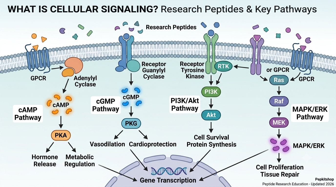
What is cellular signaling and how do research peptides interact with it and why does understanding this relationship matter for anyone working with research compounds?
Every research peptide that has been discussed in this educational series produces its biological effects through cellular signaling. A GHRP activates the ghrelin receptor. A GHRH analog activates the GHRH receptor. A melanocortin activates one of five MC receptors. A natriuretic peptide activates a guanylyl cyclase receptor. These receptor activations all produce different intracellular events. And it is those intracellular events the signaling pathways that actually create the biological outcomes researchers are studying.
Understanding what is cellular signaling and how do research peptides interact with it is the unifying framework that connects every compound education post in this series. Once you understand the five major signaling pathways that research peptides engage, the mechanisms of every compound become significantly easier to understand, remember, and apply to research design.
This guide explains what is cellular signaling from first principles, covers the five primary pathways that research peptides interact with, shows how each pathway class covered in this series maps to specific signaling architectures, addresses the emerging concept of biased signaling that the March 2026 MedUni Vienna paper has brought into sharper focus, and explains why pathway selectivity matters for research outcomes.
If you have been following this compound education series, this post is the synthesis. If you are new here, start with our post on [how peptides work in the body] first, then return here for the complete framework.
Table of Contents
What Is Cellular Signaling?
What is cellular signaling in plain scientific terms?
Once a peptide binds its receptor, it initiates a cascade of intracellular events: second messenger production such as cAMP, cGMP, and calcium ions, kinase activation including PKA, PKC, and MAPK, gene transcription changes, and ultimately altered protein synthesis and cellular behavior.
Cellular signaling is the process by which a cell receives information from its environment, transmits that information through a cascade of molecular events inside the cell, and produces a specific biological response as a consequence. It is the mechanism through which a signal originating outside a cell whether from a hormone, a peptide, a neurotransmitter, a growth factor, or any other signaling molecule produces a change in what the cell does.
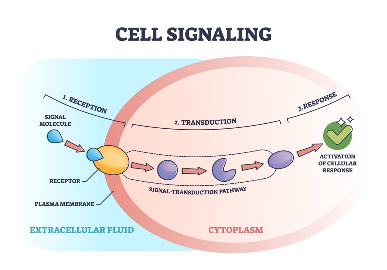
The process always follows the same general architecture regardless of which molecule is doing the signaling:
First, a signaling molecule binds to a receptor on or in the target cell. Second, the receptor undergoes a conformational change that initiates intracellular events. Third, these intracellular events are amplified through a cascade of molecular reactions. Fourth, the amplified signal reaches specific proteins that carry out the cellular response. Fifth, the signal is eventually terminated through degradation, feedback inhibition, or receptor desensitization.
What is cellular signaling in the context of research peptides is specifically the study of which receptors peptide compounds activate, which intracellular cascades those receptors initiate, and what biological outcomes those cascades produce in different cell types and tissue environments.
Research peptides represent a diverse class of short-chain amino acid sequences synthesized for laboratory investigation into cellular signaling, tissue repair, metabolic regulation, and hormone modulation. Their specificity of action and ability to finely modulate cellular signaling pathways make them valuable tools for understanding fundamental biological mechanisms.
The 4 Requirements for Any Cellular Signaling Event
What is cellular signaling built on at its most basic structural level?
Every cellular signaling event requires four components to function:
The 4 Requirements for Cellular Signaling
| Component | What It Is | Example in Peptide Biology |
|---|---|---|
| Signal | The molecule carrying information | Alpha-MSH, GHRP-6, ANP, CJC-1295 |
| Receptor | The protein that recognizes and binds the signal | MC4R, GHS-R1a, NPR-A, GHRH-R |
| Transducer | The intracellular machinery that converts receptor binding into downstream events | G-proteins, guanylyl cyclases, beta-arrestins |
| Effector | The protein that carries out the final cellular response | Adenylyl cyclase, phospholipase C, kinases |
The specificity of what is cellular signaling comes from the receptor. A peptide can only produce its biological effect in cells that carry the receptor it binds. This receptor specificity is what makes peptide-mediated signaling inherently targeted rather than generalized. Alpha-MSH cannot activate hypothalamic neurons that lack MC4R. GHRP-6 cannot stimulate GH release from cells that lack GHS-R1a. The receptor distribution across tissues is what determines where a peptide’s effects are felt.
The amplification that occurs between receptor binding and cellular response is what makes signaling so powerful relative to the tiny concentrations at which peptides operate. A single receptor binding event can activate hundreds of G-proteins, each of which can stimulate hundreds of adenylyl cyclase molecules, each of which can produce thousands of cAMP molecules, each of which can activate PKA enzymes, each of which can phosphorylate hundreds of substrate proteins. This cascade amplification produces measurable cellular changes from vanishingly small concentrations of the original signal.
The 5 Major Pathways Research Peptides Interact With
What is cellular signaling through the five primary pathways that research peptides engage?
The major signaling pathways implicated in mechanistic studies of bioactive peptides include the PI3K/Akt, MAPK/ERK, and Wnt/beta-catenin pathways among others. Peptides exert their functions by modulating key cellular signaling pathways, thereby regulating diverse biological processes related to cell proliferation.
The 5 Major Signaling Pathways in Research Peptide Biology
| Pathway | Primary Second Messenger | Key Kinase | Receptor Type | Primary Peptide Classes |
|---|---|---|---|---|
| cAMP-PKA | Cyclic AMP | Protein Kinase A | Gs-coupled GPCR | GHRH analogs, melanocortins (MC1R, MC3R, MC4R, MC5R), glucagon-like peptides |
| cGMP-PKG | Cyclic GMP | Protein Kinase G | Receptor guanylyl cyclase | Natriuretic peptides, nitric oxide-related compounds |
| Calcium-PKC | Intracellular calcium | Protein Kinase C | Gq-coupled GPCR | GHRPs through GHS-R1a, some neuropeptides |
| MAPK-ERK | No classical second messenger | ERK1/2 kinases | Multiple, often downstream of GPCRs and receptor tyrosine kinases | Tissue repair peptides, growth factors, some BPC-157 mechanisms |
| PI3K-Akt | PIP3 | Akt (PKB) | Receptor tyrosine kinases, downstream of some GPCRs | Growth factor-related peptides, insulin-like signaling, survival signaling |
These five pathways are not mutually exclusive. Many receptors activate more than one pathway simultaneously, and downstream crosstalk between pathways means that activating one pathway can influence the activity of others. Understanding what is cellular signaling in its full complexity requires recognizing that real biological responses are rarely the product of a single linear pathway but of multiple interacting signaling networks.
Pathway 1: The cAMP-PKA Pathway
What is cellular signaling through the cAMP-PKA pathway and which research peptides use it?
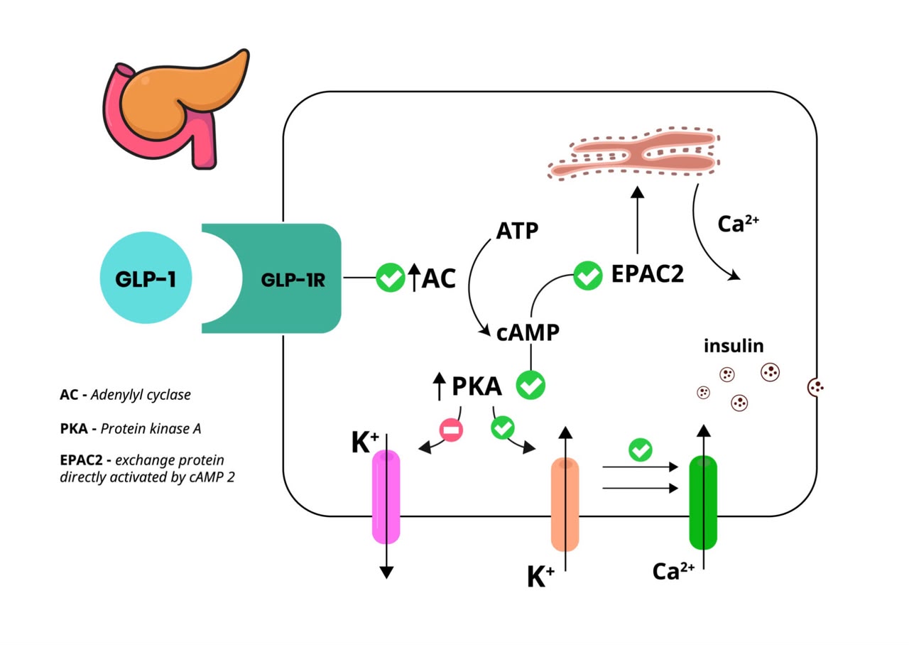
The cAMP-PKA pathway is the most widely used signaling mechanism in the peptide hormones covered in this series. It is activated by Gs-coupled G-protein coupled receptors.
When a peptide binds a Gs-coupled receptor, the receptor activates a stimulatory G-protein (Gαs). Gαs stimulates adenylyl cyclase, an enzyme in the cell membrane that converts ATP to cyclic AMP (cAMP). cAMP binds to and activates protein kinase A (PKA), which then phosphorylates numerous downstream targets including transcription factors, ion channels, and metabolic enzymes.
cAMP-PKA Pathway: Research Peptide Examples
| Peptide Class | Receptor | Pathway Outcome |
|---|---|---|
| GHRH analogs (CJC-1295, sermorelin, tesamorelin) | GHRH-R | cAMP-PKA drives GH synthesis and release from pituitary somatotrophs |
| Melanocortins (alpha-MSH, ACTH) | MC1R, MC3R, MC4R, MC5R | cAMP-PKA drives melanin synthesis (MC1R), cortisol production (MC2R), appetite suppression (MC4R) |
| GLP-1 peptides | GLP-1R | cAMP-PKA drives insulin secretion from pancreatic beta cells |
| Glucagon | Glucagon receptor | cAMP-PKA drives hepatic glucose production |
| VIP (vasoactive intestinal peptide) | VPAC receptors | cAMP-PKA drives smooth muscle relaxation, immune modulation |
The cAMP-PKA pathway not only regulates hormone secretion but also exerts potent anti-apoptotic effects in cardiovascular and neural tissues, promoting cell survival through multiple downstream mechanisms.
The cAMP pathway has two primary biological outputs depending on the cell type. In secretory cells like pituitary somatotrophs and pancreatic beta cells, cAMP-PKA drives vesicle fusion and hormone secretion. In other cell types, cAMP-PKA activates the transcription factor CREB, which drives gene expression changes that produce longer-term cellular adaptations.
Pathway 2: The cGMP-PKG Pathway
What is cellular signaling through the cGMP-PKG pathway and which research peptides use it?
The cGMP-PKG pathway is distinct from the cAMP pathway in its receptor mechanism. While cAMP is produced by adenylyl cyclase activated indirectly through G-proteins, cGMP can be produced by two different mechanisms: through soluble guanylyl cyclase activated by nitric oxide, or through membrane-bound guanylyl cyclase receptors activated by natriuretic peptides.
As covered in our post on [what is a natriuretic peptide], the natriuretic peptide receptors NPR-A and NPR-B are themselves guanylyl cyclase enzymes. When ANP, BNP, or CNP bind their extracellular domains, the intracellular guanylyl cyclase domain is directly activated, producing cGMP without any G-protein intermediary. This makes the cGMP-PKG pathway mechanistically distinct from the cAMP-PKA pathway despite producing similar downstream amplification.
cGMP-PKG Pathway: Key Features
| Feature | cGMP-PKG via Natriuretic Peptides | cGMP via Nitric Oxide |
|---|---|---|
| Second messenger source | Membrane bound guanylyl cyclase receptor | Soluble guanylyl cyclase activated by NO |
| Receptor type | Receptor guanylyl cyclase (NPR-A, NPR-B) | Intracellular enzyme, not a surface receptor |
| Initiating signal | Peptide hormone binding (ANP, BNP, CNP) | Nitric oxide gas diffusion |
| cGMP produced | Yes, directly by receptor | Yes, by soluble guanylyl cyclase |
| PKG activation | Yes | Yes |
| Primary outcomes | Natriuresis, vasodilation, cardioprotection | Vasodilation, platelet inhibition, neurotransmission |
| Interaction | Both pathways feed into same cGMP pool | Additive cardiovascular effects |
The convergence of natriuretic peptide signaling and nitric oxide signaling on the same cGMP-PKG pathway explains the complementary cardiovascular effects of these two systems and why conditions that impair both simultaneously (like endothelial dysfunction) produce more severe cardiovascular consequences than impairment of either alone.
Pathway 3: The Calcium-PKC Pathway
What is cellular signaling through the calcium-PKC pathway and which research peptides use it?
The calcium-PKC pathway is activated by Gq-coupled G-protein coupled receptors. When a peptide binds a Gq-coupled receptor, the activated Gαq stimulates phospholipase C (PLC). PLC cleaves a membrane phospholipid into two second messengers simultaneously: inositol trisphosphate (IP3) and diacylglycerol (DAG).
IP3 travels to the endoplasmic reticulum where it triggers release of stored calcium ions into the cytoplasm. The elevated intracellular calcium, combined with DAG, activates protein kinase C (PKC). PKC then phosphorylates downstream targets that produce the cellular response.
Calcium-PKC Pathway: Research Peptide Examples
| Peptide Class | Receptor | Pathway Outcome |
|---|---|---|
| GHRPs (GHRP-6, GHRP-2, hexarelin) | GHS-R1a | Calcium-PKC drives GH secretion from pituitary somatotrophs, appetite signaling in hypothalamus |
| Oxytocin | Oxytocin receptor | Calcium-PKC drives uterine contraction, social bonding neurotransmission |
| Vasopressin (ADH) | V1 receptor | Calcium-PKC drives vasoconstriction |
| Endothelin | ET-A, ET-B receptors | Calcium-PKC drives vascular smooth muscle contraction |
| Substance P | NK1 receptor | Calcium-PKC drives pain and inflammatory signaling |
The calcium-PKC pathway produces different biological outcomes than the cAMP-PKA pathway even when both are activated in the same cell type. As covered in our post on [what is a growth hormone releasing peptide], GHRPs activate GHS-R1a through the calcium-PKC pathway, while GHRH analogs activate the GHRH receptor through the cAMP-PKA pathway. Both ultimately drive GH release from pituitary somatotrophs, but through distinct intracellular mechanisms that produce the synergistic amplification when both pathways are activated simultaneously.
This mechanistic distinction is also why GHRPs and GHRH analogs have different secondary effect profiles beyond GH release they are activating fundamentally different signaling cascades that have different downstream targets in cells throughout the body.
Pathway 4: The MAPK-ERK Pathway
What is cellular signaling through the MAPK-ERK pathway and how do research peptides interact with it?
The MAPK-ERK pathway, where MAPK stands for mitogen-activated protein kinase and ERK stands for extracellular signal-regulated kinase, is a central pathway for cell proliferation, differentiation, survival, and tissue repair responses.
The MAPK/ERK pathway is one of the major signaling pathways implicated in mechanistic studies of bioactive peptides promoting cell proliferation. Peptides promote the proliferation of macrophages and lymphocytes via the NF-kB and MAPK signaling pathways, and also reduce the secretion of pro-inflammatory cytokines like TNF-alpha, IL-6, NO, and PGE2, thereby offering anti-inflammatory and immunomodulatory effects.
Unlike the cAMP-PKA and cGMP-PKG pathways, MAPK-ERK is not activated directly by a single receptor class. It can be activated downstream of receptor tyrosine kinases, GPCRs, and multiple other receptor types through various adapter protein cascades. The core cascade involves sequential phosphorylation: a signal activates Ras, which activates Raf, which activates MEK, which activates ERK1/2. Activated ERK1/2 then enters the nucleus and phosphorylates transcription factors that drive gene expression changes.
MAPK-ERK Pathway in Research Peptide Contexts
| Research Context | How MAPK-ERK Is Engaged | Biological Outcome |
|---|---|---|
| Tissue repair peptides | Receptor interactions activate Ras-Raf-MEK-ERK cascade | Cell proliferation, fibroblast activation, collagen synthesis |
| GHRH analog extrapituitary effects | PI3K-Akt activation of downstream MAPK-ERK | Cardiovascular cell survival and proliferation |
| Growth factor signaling | Receptor tyrosine kinase activation of Ras | Muscle satellite cell proliferation, tissue regeneration |
| Anti-inflammatory peptide effects | Inhibition of MAPK pathway branches | Reduced cytokine production, immune cell modulation |
| Neuroprotective peptides | ERK activation promotes neuronal survival | Anti-apoptotic signaling in neural tissue |
The MAPK-ERK pathway is particularly relevant to understanding tissue repair peptide biology. Many of the research mechanisms documented for compounds studied in recovery and regeneration contexts involve ERK1/2 activation driving fibroblast proliferation, collagen synthesis, and cellular migration the core processes of tissue repair. Compounds that modulate the FAK-paxillin pathway mentioned in growth factor research also ultimately converge on MAPK signaling through effects on cell migration and adhesion.
Pathway 5: The PI3K-Akt Pathway
What is cellular signaling through the PI3K-Akt pathway and how does it appear in research peptide biology?
The PI3K-Akt pathway, where PI3K stands for phosphoinositide 3-kinase and Akt is also known as protein kinase B, is a central regulator of cell survival, metabolism, growth, and proliferation. It is one of the most frequently dysregulated pathways in cancer biology and one of the most important for understanding survival signaling in cardiovascular and neural tissue.
PI3K is activated by receptor tyrosine kinases (such as the insulin receptor and IGF-1 receptor), by some GPCRs through beta-gamma G-protein subunits, and by RAS signaling. When activated, PI3K phosphorylates a membrane phospholipid called PIP2 into PIP3. PIP3 recruits Akt to the membrane where it is phosphorylated and activated. Active Akt then phosphorylates numerous downstream targets that collectively promote cell survival, protein synthesis, and metabolic regulation.
PI3K-Akt Pathway in Research Peptide Biology
| Peptide Context | PI3K-Akt Role | Biological Consequence |
|---|---|---|
| IGF-1 signaling downstream of GH secretagogues | IGF-1 receptor activates PI3K-Akt | Muscle protein synthesis, cell survival, anti-apoptosis |
| GHRH analog extrapituitary effects | GHRH-R activates PI3K-Akt in cardiac and neural tissue | Cardioprotection, neuroprotection independent of GH |
| Insulin-related peptides | Insulin receptor activates PI3K-Akt | Glucose uptake, glycogen synthesis, anti-apoptosis |
| Growth factor releasing peptides | VEGF receptor activates PI3K-Akt in endothelial cells | Angiogenesis, endothelial cell survival |
| Tissue repair contexts | Multiple receptor inputs converge on PI3K-Akt | Cell survival during repair, fibroblast proliferation |
GHRH analog mediated cardiovascular protection occurs through PI3K-Akt signaling promoting cell survival and endothelial nitric oxide production. These benefits occur independently of GH and IGF-1 signaling, highlighting their therapeutic potential for cardiovascular and cerebrovascular diseases independently of the GH axis.
The finding that GHRH analog cardiovascular protection operates through direct PI3K-Akt signaling in cardiac tissue, independent of GH and IGF-1, illustrates a critical point about what is cellular signaling in research peptide contexts. The same peptide can activate different signaling pathways in different cell types, producing effects that have no relationship to the pathway we originally think of as primary. Researchers who study only the pituitary GH-releasing function of GHRH analogs miss the direct cardiovascular effects that operate through a completely separate signaling architecture.
How the Compound Education Series Maps to These Pathways
What is cellular signaling showing us when we map everything covered in this series?
Complete Pathway Map: Pepkitshop Compound Education Series
| Compound Class | Primary Receptor | Primary Pathway | Secondary Pathways |
|---|---|---|---|
| GHRPs | GHS-R1a (ghrelin receptor) | Calcium-PKC (Gq-coupled) | MAPK-ERK downstream, anti-inflammatory NF-kB inhibition |
| GHRH analogs | GHRH-R | cAMP-PKA (Gs-coupled) | PI3K-Akt and MAPK-ERK in extrapituitary tissues |
| Melanocortins | MC1R, MC3R, MC4R, MC5R | cAMP-PKA (Gs-coupled) | Kir7.1 ion channel (MC4R independent of cAMP) |
| Natriuretic peptides | NPR-A (ANP, BNP), NPR-B (CNP) | cGMP-PKG (receptor guanylyl cyclase) | Gi-coupled via NPR-C |
| GLP-1 class | GLP-1R | cAMP-PKA (Gs-coupled) | PI3K-Akt in beta cells |
| IGF-1 | IGF-1R (receptor tyrosine kinase) | PI3K-Akt, MAPK-ERK | Multiple downstream cascades |
| Tissue repair peptides | Multiple: VEGFR2, growth factor receptors | MAPK-ERK, angiogenesis pathways | PI3K-Akt, nitric oxide-cGMP |
Reading this table shows clearly that what is cellular signaling in research peptide biology is not a monolithic mechanism but a diverse ecosystem of receptor-pathway combinations. GHRPs and GHRH analogs both drive GH release but through calcium-PKC and cAMP-PKA respectively. Melanocortins and GHRH analogs both use cAMP-PKA but through entirely different receptors with different tissue distributions. Natriuretic peptides use cGMP-PKG through a mechanism that bypasses G-proteins entirely.
This pathway diversity is also why combining research compounds from different pathway classes produces effects that are not simply additive. GHRP plus GHRH analog synergy exists precisely because two independent signaling pathways are simultaneously activated, both converging on the same final output of GH release through mechanistically distinct routes.
Biased Signaling: The March 2026 MedUni Vienna Discovery
What is cellular signaling showing in the most current 2026 research?
A research team led by Christian Gruber from the Center for Physiology and Pharmacology at MedUni Vienna highlighted a promising new approach to drug discovery focused on the targeted modulation of certain intracellular signaling proteins as a strategy for controlling disease-relevant signaling pathways and reducing side effects. The focus is on beta-arrestins, multifunctional switching points in cellular signal transduction. These proteins regulate, amplify or direct signals within the cell. The team showed how tailor-made peptides, generated by computational design or derived from chemical libraries, bind specifically to target structures such as receptors or arrestins. The findings are published in the journal Trends in Pharmacological Sciences, DOI 10.1016/j.tips.2026.01.004.
This finding from MedUni Vienna introduces a concept called biased signaling that is increasingly important for understanding what is cellular signaling in peptide research contexts.
When most GPCRs are activated, they initially couple to G-proteins to produce the classical second messenger responses (cAMP, cGMP, calcium). After a brief period, beta-arrestins are recruited to the receptor. Beta-arrestins serve two functions: they terminate the G-protein signaling by uncoupling the receptor from its G-protein (a process called desensitization), and they initiate their own independent signaling cascade through different downstream pathways including MAPK-ERK.
G-Protein vs Beta-Arrestin Biased Signaling
| Parameter | G-Protein Signaling | Beta-Arrestin Signaling |
|---|---|---|
| Timing | Immediate, within seconds of receptor activation | Delayed, following beta-arrestin recruitment |
| Second messengers | cAMP, cGMP, calcium, DAG | No classical second messenger |
| Downstream effects | Acute cellular responses (secretion, contraction, relaxation) | Internalization, gene expression, receptor trafficking |
| Duration | Brief, terminated by desensitization | More prolonged, continues after G-protein uncoupling |
| Therapeutic relevance | Targets for most existing drugs | Emerging target class for biased agonist design |
| Research peptide relevance | Primary mechanism for most peptide classes studied | Increasingly recognized as parallel signaling pathway |
Biased agonism refers to the ability of some ligands to preferentially activate either G-protein signaling or beta-arrestin signaling at the same receptor. A G-protein biased agonist produces primarily the classical cAMP or calcium responses with minimal beta-arrestin signaling. A beta-arrestin biased agonist produces primarily the MAPK-ERK and internalization effects with minimal G-protein response.
The MedUni Vienna work shows that tailor-made peptides can be designed to target beta-arrestins directly as intracellular signaling proteins, rather than targeting surface receptors. This represents a conceptually new approach where the peptide acts inside the cell rather than at the cell surface raising the possibility of research compounds that bypass receptor binding entirely and directly modulate the intracellular signal transduction machinery.
Scientists are increasingly studying molecules capable of influencing multiple signaling pathways simultaneously. This approach reflects a broader shift within biological research toward understanding how biological systems coordinate responses to internal and external stimuli through interconnected signaling networks rather than linear pathways.
Why Pathway Selectivity Matters for Research Design
What is cellular signaling’s practical implication for how research peptide studies should be designed?
Understanding which signaling pathways a research peptide activates is not academic knowledge. It directly affects how experiments should be designed, how results should be interpreted, and what controls are necessary to draw valid conclusions.
Pathway Selectivity and Research Design Implications
| Research Scenario | Pathway Knowledge Required | Consequence of Ignoring It |
|---|---|---|
| Combining two peptide classes | Which pathways each activates | Misinterpreting additive as synergistic or synergistic as additive |
| Cell type selection | Which receptors and pathways are present in chosen cells | Studying wrong cell type for the pathway of interest |
| Inhibitor studies | Which specific kinases or enzymes to inhibit | Using wrong inhibitor, blocking unrelated pathway |
| Dose-response experiments | How pathway amplification affects dose-effect relationships | Misinterpreting steep dose-response curves |
| Interpretation of off-target effects | What secondary pathways the compound also activates | Attributing effects to wrong mechanism |
| Combination with pharmacological agents | Pathway crosstalk and drug-peptide interactions | Unexpected interactions between peptide and concurrent treatments |
Research peptides find applications in many areas of biomedical research. Their specificity of action and ability to finely modulate cellular signaling pathways make them valuable tools for understanding fundamental biological mechanisms. The study of tissue regeneration mechanisms, cardiac protection, neuroprotective factors, cell proliferation and apoptosis, and signaling pathways involved in myocardial repair all depend on understanding which pathways specific peptide compounds activate.
The specificity principle works in both directions. The fact that a research peptide activates a specific pathway is what makes it a useful research tool for studying that pathway. But the fact that it may also activate secondary pathways through receptor crosstalk, downstream convergence, or biased signaling mechanisms means that attributing observed effects solely to the primary pathway requires appropriate controls.
Understanding what is cellular signaling at the level of specific pathways, second messengers, and downstream kinases is therefore the foundation of rigorous research peptide science. It is what separates mechanistically informed research from empirical observation without interpretive framework.
More detail on current signaling research as it applies to specific peptide compounds is available through the National Library of Medicine and peer reviewed research databases.
Frequently Asked Questions
What is cellular signaling in simple terms?
What is cellular signaling at its most basic is the process by which information from outside a cell (in the form of a hormone, peptide, or other signaling molecule) is transmitted through a cascade of molecular events inside the cell to produce a specific biological response. Every research peptide produces its effects through cellular signaling.
What is cellular signaling and how do research peptides interact with it through receptors?
What is cellular signaling and how do research peptides interact with it at the receptor level is through binding to specific proteins on the cell surface that recognize the peptide’s molecular structure. This binding triggers the receptor to change shape, which activates intracellular signaling proteins that amplify and transmit the signal through a cascade of molecular events.
What is the difference between cAMP and cGMP signaling?
What is cellular signaling through cAMP involves Gs-coupled GPCRs activating adenylyl cyclase to produce cAMP, which activates PKA. Most peptide hormone classes including GHRH analogs and melanocortins use this pathway. What is cellular signaling through cGMP involves either natriuretic peptide receptors that are themselves guanylyl cyclase enzymes, or soluble guanylyl cyclase activated by nitric oxide. cGMP activates PKG rather than PKA, producing different downstream effects.
What is cellular signaling and how do research peptides interact with it through MAPK-ERK?
What is cellular signaling through MAPK-ERK is a pathway activated downstream of multiple receptor types that drives cell proliferation, differentiation, and survival. Research peptides interact with this pathway both directly through receptors that couple to MAPK cascades and indirectly through downstream convergence from primary signaling pathways like cAMP-PKA and PI3K-Akt.
What is biased signaling and why does it matter for research peptides?
Biased signaling refers to the ability of some ligands to preferentially activate either G-protein signaling or beta-arrestin signaling at the same receptor. The March 2026 MedUni Vienna paper expanded this concept by showing that tailor-made peptides can target beta-arrestins directly as intracellular signaling proteins, bypassing surface receptor binding entirely. This matters for research peptides because different structural variants of the same peptide may have different G-protein versus beta-arrestin bias ratios, producing different effect profiles even at the same receptor.
Why do GHRPs and GHRH analogs produce synergistic rather than simply additive GH release?
What is cellular signaling explaining about this synergy is that GHRPs activate calcium-PKC through the ghrelin receptor while GHRH analogs activate cAMP-PKA through the GHRH receptor. These are independent intracellular cascades that both converge on GH secretion from pituitary somatotrophs through different mechanisms. Simultaneous activation of two independent pathways produces synergistic rather than additive output because the mechanisms address different rate-limiting steps in GH release simultaneously.
What is cellular signaling and how do research peptides interact with it at the gene expression level?
What is cellular signaling ultimately producing in many cases is changes in gene expression. PKA, PKG, PKC, ERK1/2, and Akt all phosphorylate transcription factors that alter which genes are being transcribed. This means research peptides that activate signaling pathways ultimately change the pattern of proteins being produced by target cells, with effects that can persist long after the peptide itself has been degraded.
Key Takeaways
- What is cellular signaling and how do research peptides interact with it is the unifying framework connecting every compound class covered in this educational series
- Cellular signaling requires four components: the signal (peptide), the receptor, the transducer (G-protein or guanylyl cyclase), and the effector (kinase or other downstream protein)
- Five major pathways are engaged by research peptides: cAMP-PKA (activated by GHRH analogs and melanocortins through Gs-coupled GPCRs), cGMP-PKG (activated by natriuretic peptides through receptor guanylyl cyclases), calcium-PKC (activated by GHRPs through Gq-coupled GHS-R1a), MAPK-ERK (activated by multiple receptor types, central to tissue repair signaling), and PI3K-Akt (activated by receptor tyrosine kinases and downstream of some GPCRs)
- GHRPs and GHRH analogs use different pathways — calcium-PKC and cAMP-PKA respectively — which explains the mechanistic basis for their synergistic GH release when combined
- Natriuretic peptides use a receptor mechanism fundamentally different from GPCRs: their receptors are themselves guanylyl cyclase enzymes that directly produce cGMP without G-protein intermediation
- The March 2026 MedUni Vienna paper published in Trends in Pharmacological Sciences identified beta-arrestins as a novel intracellular target for tailor-made peptides, expanding the concept of cellular signaling beyond surface receptor binding to direct intracellular signaling protein modulation
- Pathway selectivity matters for research design: the pathways a compound activates determine which cell types to study, which inhibitors to use as controls, and how to interpret effects that may involve multiple converging mechanisms
Continue Learning
This post synthesizes the compound education series from Pepkitshop. Complete related reading:
- [How Do Peptides Work in the Body?] The foundational guide to receptor binding, second messengers, and the four modes of cellular communication
- [What Is a Growth Hormone Releasing Peptide?] The GHRP class and calcium-PKC signaling through GHS-R1a
- [What Is a GHRH Analog?] The GHRH analog class and cAMP-PKA signaling through the GHRH receptor
- [What Is a Melanocortin Peptide?] The MC receptor family and cAMP-PKA signaling across five receptor subtypes
- [What Is a Natriuretic Peptide?] The natriuretic peptide family and cGMP-PKG signaling through receptor guanylyl cyclases
The complete Pepkitshop educational library is available at pepkitshop.com. Evidence based peptide science explained clearly without hype.
This content is for educational and informational purposes only. It does not constitute medical advice, diagnosis, or treatment recommendations. Research peptides are for laboratory use only and are not approved for human consumption or therapeutic use. Always consult a qualified healthcare professional before making any decisions related to your health.